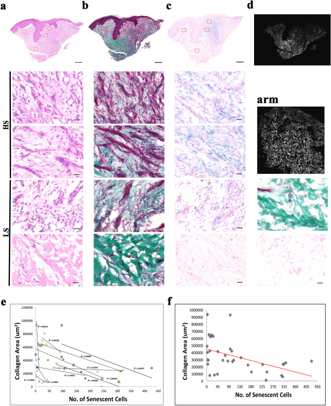
Figure 1.
Staining of nuclei, collagen and senescent cells of venous leg ulcer biopsies revealed a strong link between presence of senescent cells and collagen area. (a) Haematoxylin and eosin staining of nuclei (purple) and extracellular proteins (pink) in a venous leg ulcer. Montage of high-power images [1200 (W) × 1000 (H) μm] of an 8 μM cryosection thick—two from high senescence (HS) regions and two from low senescence (LS) regions of a venous leg ulcer taken using × 20 objective lens. (b) MT staining collagen (green), muscle and keratin (red) and cytoplasm (pink/red) in a sister section. Montage followed by (c) X-gal staining as a marker for senescent cells (senescent cells (blue), nuclei and cytoplasm (pink). Scale bar: 500 μm (montage) and 20 μm (high power). (d) SHG imaging of collagen (white) in × 20 for VLU and arm tissue. Scale bar: 200 μm. MT and X-gal images of the arm tissue are also shown. (e) Regression plots of collagen area (x-axis) against senescent cell number (y-axis) of individual patients of the VLU cohort. (f) Single regression plot of the VLU cohort.

