FIGURE 3.
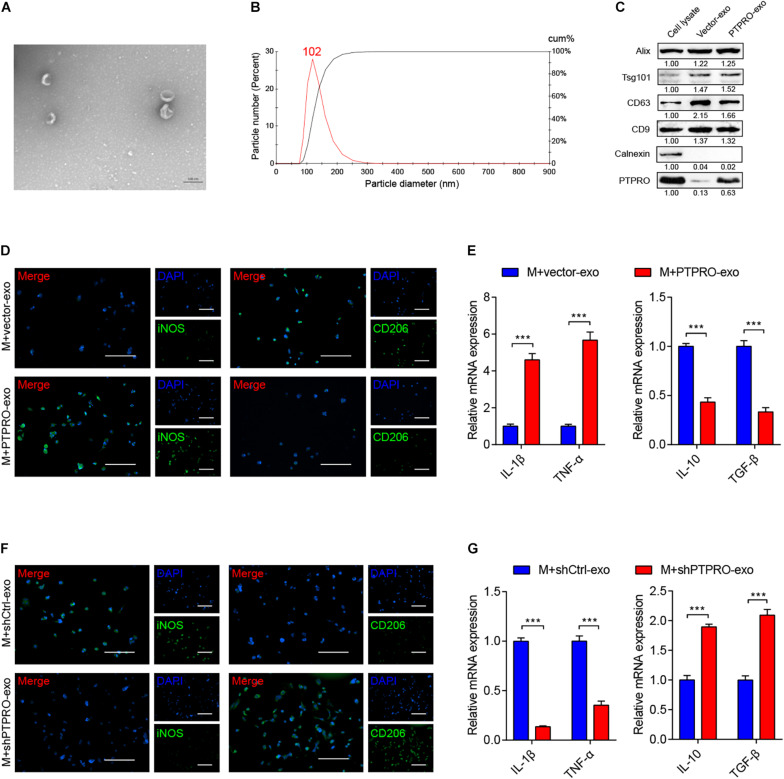
Tumor cell-derived PTPRO inflected macrophage polarization via exosome carrier. (A) Electron microscopy images of exosomes isolated from ZR-75-1-PTPRO cells. (B) Nanoparticle tracking analysis (NTA) of exosomes isolated from ZR-75-1-PTPRO cells. (C) The expressions of Alix, TSG101, CD63, CD9, Calnexin, and PTPRO were measured by immunoblotting in exosomes isolated from ZR-75-1-PTPRO cells. Relative protein expression was quantified. (D) Representative images of immunofluorescence staining of iNOS and CD206 in THP1-derived macrophages after treating with ZR-75-1-vector-exo or ZR-75-1-PTPRO-exo for 48 h. Scale bars = 50 μm. (E) qRT-PCR was applied to detect the relative expression of IL-1β, TNF-α, IL-10, and TGF-β in THP-1 derived macrophage after treating with ZR-75-1-vector-exo or ZR-75-1-PTPRO-exo for 48 h. (F) Representative images of immunofluorescence staining of iNOS and CD206 in THP1-derived macrophages after treating with ZR-75-1-shCtrl-exo or ZR-75-1-shPTPRO-exo for 48 h. Scale bars = 50 μm. (G) qRT-PCR was applied to detect the relative expression of IL-1β, TNF-α, IL-10, and TGF-β in THP-1 derived macrophage after treating with ZR-75-1-shCtrl-exo or ZR-75-1-shPTPRO-exo for 48 h. Error bars, SEM. ***P < 0.001 by Student’s t-test.

