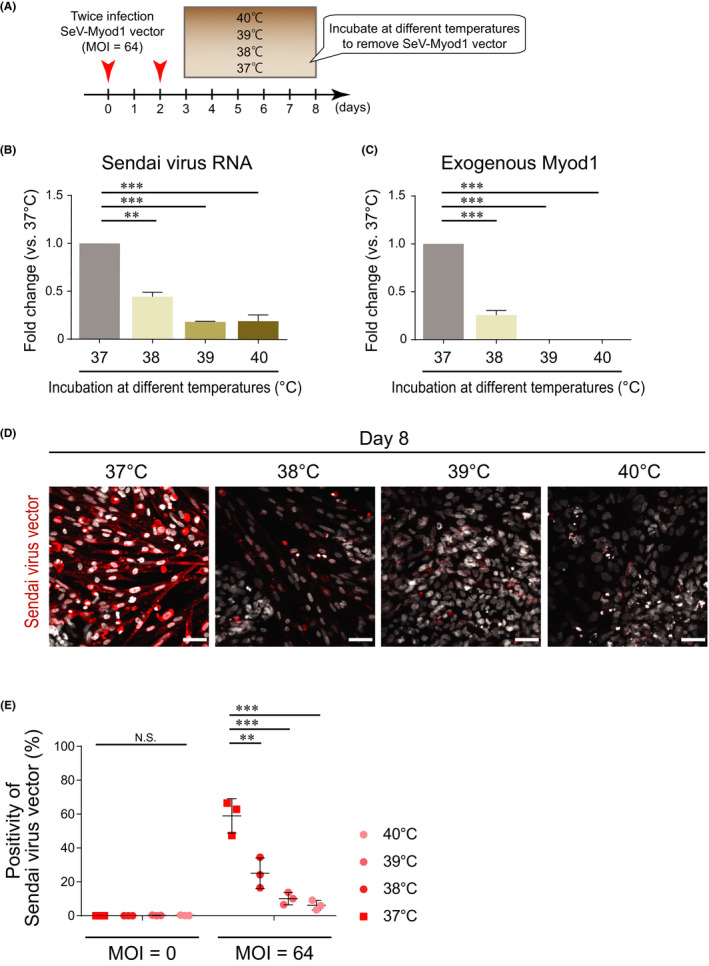
FIGURE 2.

Transient cultivation at high temperature removed infected SeV‐Myod1. (A) Schema of SeV‐Myod1 vector infection and culture condition. The cells were incubated at 37ºC, 38ºC, 39ºC or 40ºC from day 3 to day 8, and the removal of SeV‐Myod1 vector was evaluated. (B) SeV RNA was analysed on day 8 and compared among the different temperature conditions. (Independent experiments n = 3, mean ± SD, **p < 0.01, ***p < 0.001). (C) The mRNA level of exogenous Myod1 delivered by SeV‐Myod1 was analysed on day 8 and compared among the different temperature conditions. (Independent experiments n = 3, mean ± SD, ***p < 0.001). (D) Differentiating cells were immunostained for SeV antibody (red). Nuclei were stained with DAPI (white). Scale bar = 100 μm. (E) Positivity of Sendai virus vector against total DAPI (%) was quantified and compared among the different temperature conditions. (Independent experiments, n = 3, mean ± SD, **p < 0.01, ***p < 0.001, NS: not significant)
