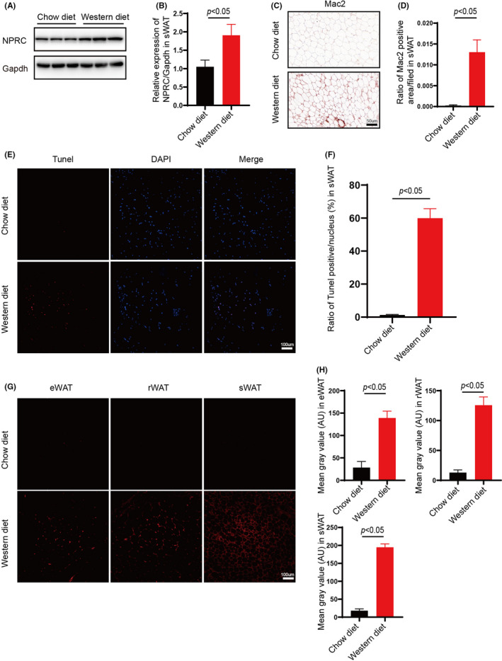
FIGURE 1.

Increased NPRC expression and inflammation in white adipose tissue of western‐diet ApoE knockout hypercholesterolemic mice. (A) NPRC expression in subdermal adipose tissue of ApoE−/− mice fed with chow diet and western diet measured by Western blot. (B) Quantification of relative expression in subdermal adipose tissue of NPRC in ApoE−/−mice fed with chow diet and western diet (n = 5). (C) NPRC in subdermal adipose tissue from ApoE−/− mice fed with chow diet and western diet stained by immunohistochemistry. (D) Quantification of NPRC in subdermal adipose tissue from ApoE−/− mice fed with chow diet and western diet stained by immunohistochemistry (n = 5). (E) Apoptosis assessed in subdermal adipose tissue from ApoE−/− mice fed with chow diet and western diet measured by TUNEL kit (n = 5). WT and NPRC−/− mice were measured by Western blot. (F) Quantification of apoptosis assessed in subdermal adipose tissue from ApoE−/− mice fed with chow diet and western diet (n = 5). (G) ROS assessed in white adipose tissue from ApoE−/− mice fed with chow diet and western diet measured by DHE staining. (H) Quantification of ROS assessed in white adipose tissue from ApoE−/− mice fed with chow diet and western diet (n = 5)
