
FIGURE 4.
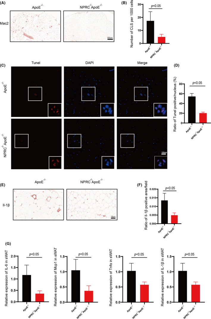
Loss of NPRC decreases white adipose inflammation and apoptosis of adipocytes in hypercholesterolemic mice. (A) The representative images of immunohistochemical staining for Mac2 in subdermal white adipose tissue. (B) Quantification of CLS density positive area (n = 5). (C) The representative images of TUNEL assay for subdermal white adipose tissue in APOE−/−NPRC−/− mice and APOE−/− mice. (D) Quantification of ratio of TUNEL positive pots in subdermal white adipose tissue in APOE−/−NPRC−/− mice and APOE−/− mice (n = 5). (E) The representative images of immunohistochemical staining for IL‐1β in subdermal white adipose tissue. (F) Quantification of ratio of IL‐1β positive area (n = 5). (G) IL‐6, Mcp1, TNFα and IL‐1β in ApoE−/−NPRC−/− mice and ApoE−/− mice measured by qPCR (n = 5)