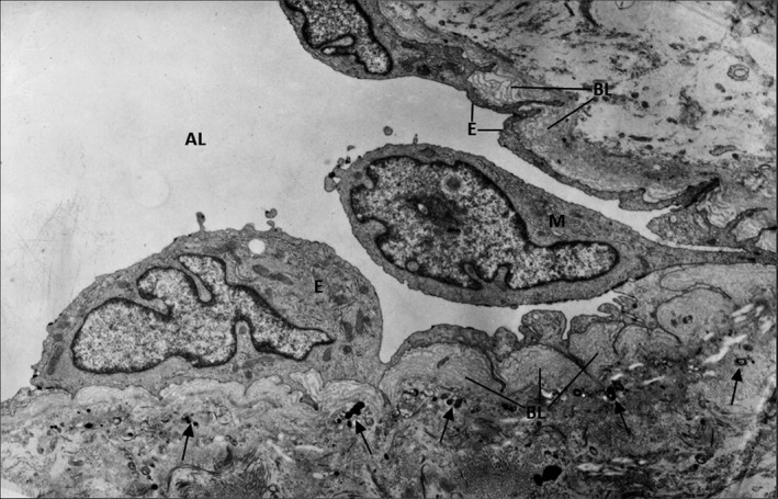
FIGURE 2.

Early‐stage ultrastructural modifications of the aortic valve lesion occurred in a hyperlipemic/diabetic hamsters. Under a continuous endothelium (E) having thin areas intercalated within zones in which the cell is highly enriched in biosynthetic organelles, there is a characteristic hyperplasic, multilayered basal lamina (BL). The proliferated matrix contains numerous calcification cores (arrow). A plasma monocyte (M) insinuates a pseudopod between two valvular endothelial cells. (AL), aortic lumen. x7000. By permission from American Journal of Pathology, 148, 3, 1996, p. 1004, Figure 8
