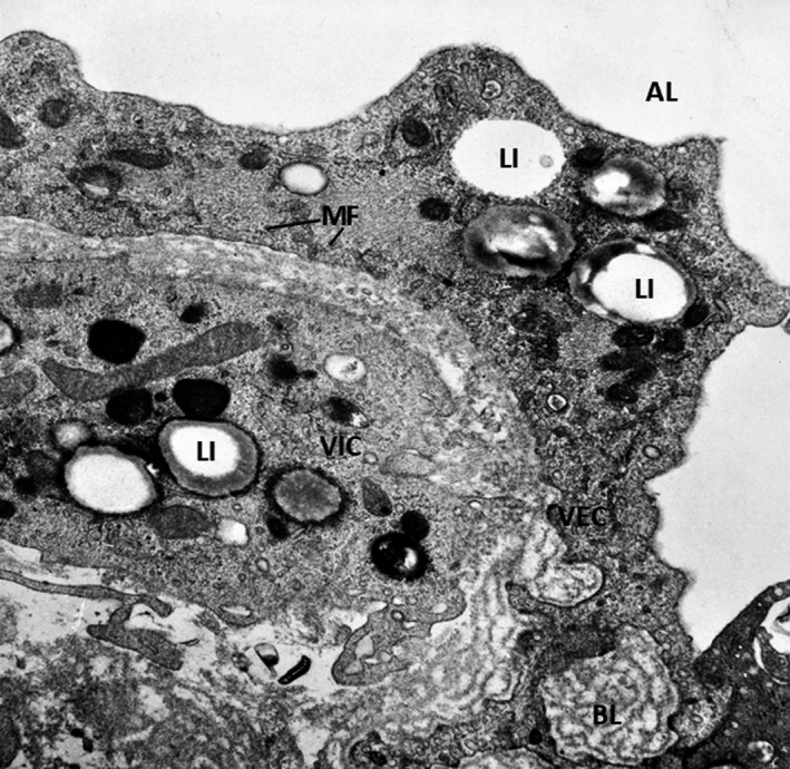
FIGURE 3.

Ultrastructure of a lesion of the aortic valve in experimental hyperglycemia/hyperlipemia (4 weeks). The pathology progresses rapidly and the alterations include thickened VECs rich in organelles, microfilaments (MF), cytoplasmic lipid inclusions (LI), a multilayered basal lamina (BL) and the presence of valvular interstitial cell (VIC)‐ containing numerous lipid inclusions. X 24,000. By permission from American Journal of Pathology, 148, 3, 1996, p.1005, Figure 9
