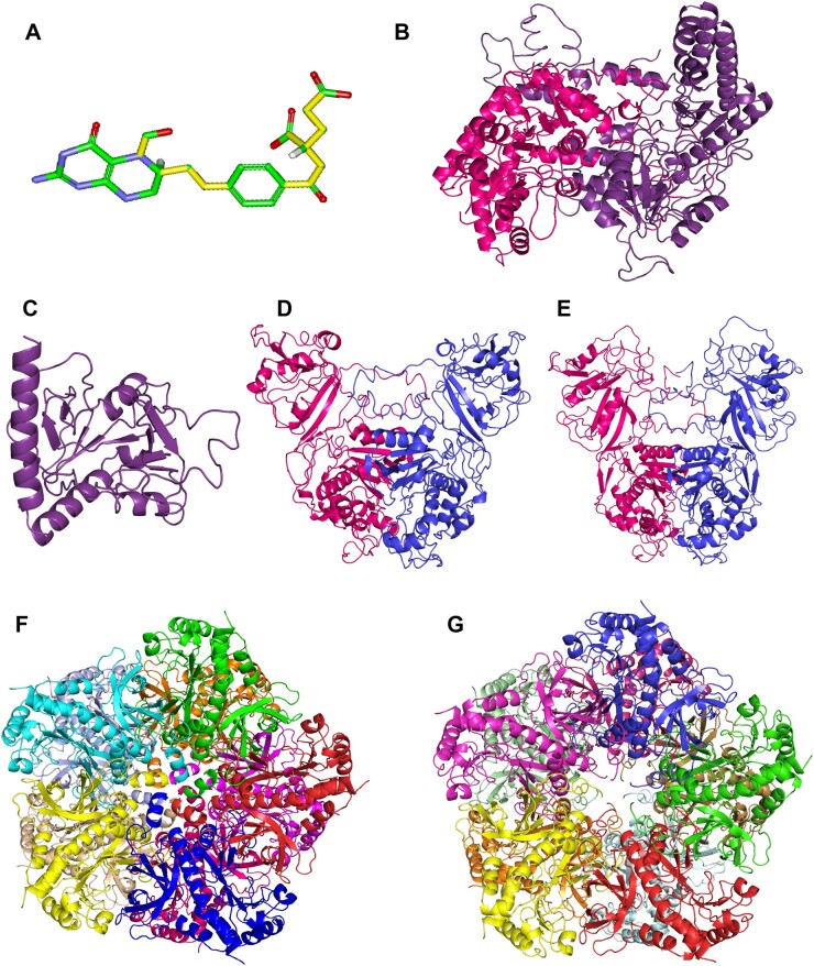
Figure 5.
Structure of the ligand 5-F-THF and homology models of the selected FFBPs. 3D structure of 5-F-THF (A) and homology models of the two known FFBPs—SHMT1 (B) and 5FCL (C), and four high-affinity FFBPs –AtDHFR-TS1 (D), AtDHFR-TS2 (E), AtGLN1;1 (F) and AtGLN1;4 (G). The 5-F-THF molecule shown in stick model and its 11 rotatable bonds are presented in yellow (color of atoms: green for C, red for oxygen, blue for N, and white for hydrogen).

