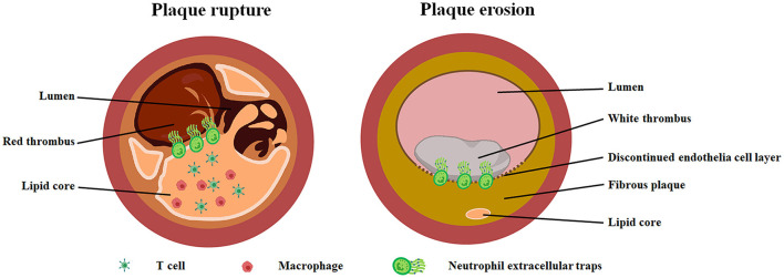
Figure 1.
Pathological characteristics of plaque rupture and plaque erosion. Ruptured plaque (left image) is featured with a larger lipid core containing abundant macrophages and T cells. Red thrombus was observed in the small lumen. Eroded plaque (right image) has a large lumen with white thrombus and fibrous plaque tissue characterized by little or no lipid deposition. In particularly, there is discontinuous endothelial cell layer in eroded plaque. Neutrophil extracellular traps was found at the junction of plaque tissue and thrombus in both eroded and ruptured plaque.

