
Figure 7.
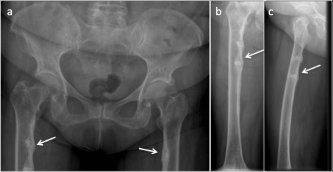
(a) Anteroposterior radiograph of the pelvis. (b) Anteroposterior and (c) Lateral radiograph of the right femur showing looser zones of osteomalacia. Multiple, bilateral lucency which involve only a part of femoral shaft along the medial aspect, oriented perpendicular to the cortex with associated surrounding sclerosis.