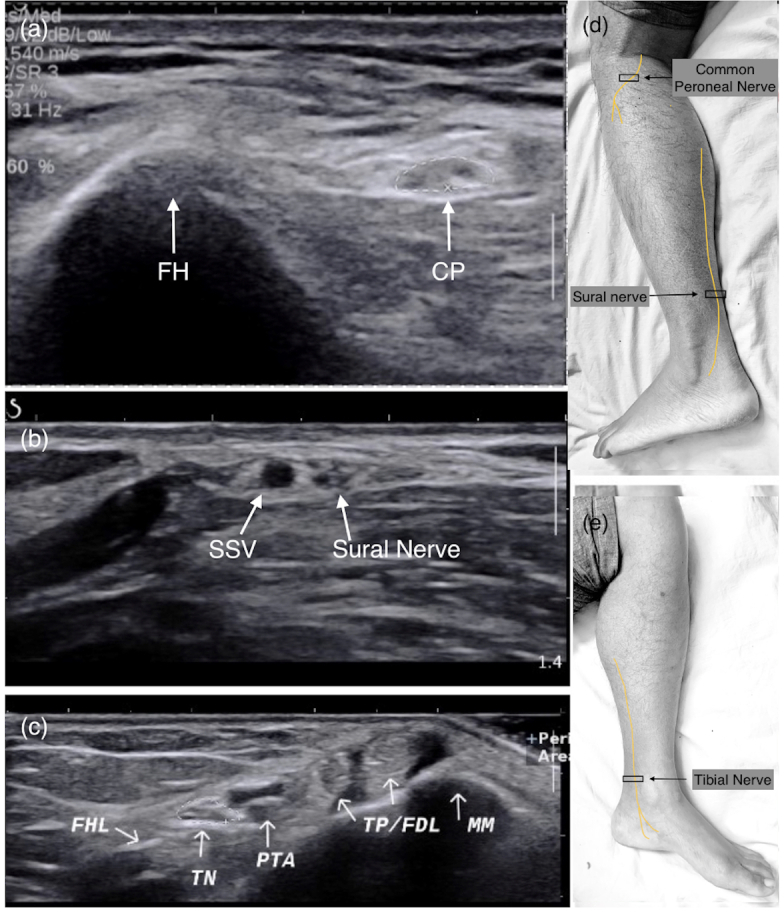
Figure 2.

Transverse ultrasound section of common peroneal, sural and tibial nerves (marked with white arrows) at different locations with anatomical landmarks. (a) CP nerve near the FH. (b) Sural nerve distal leg adjacent to SSV. (c) TN at ankle (MM, TP/FDL, PTA and veins, FHL tendon). (d, e) Image showing course of nerve with probe position (black box). CP, common peroneal; FDL, flexor tendons; FH, fibular head; FHL, flexor hallucis longus; MM, medial malleolus; PTA, posterior tibial artery; SSV, short saphenous vein; TN, tibial nerve.
