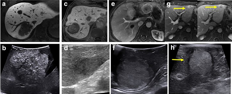
Figure 2.
MRI and corresponding IOUS images of a spectrum of liver lesions. Mucinous CRC metastatic lesion on hepatobiliary phase MRI (a) demonstrates fine calcifications and a hyperechoic appearance at IOUS (b), while a different 88-year-old male with multifocal CRC on hepatobiliary phase MRI (c) demonstrates a lobulated ill-defined hypoechoic lesion at IOUS (d). A heterogeneously enhancing lesion on arterial phase MRI imaging (e) found to be metastatic NET in a 57-year-old female was hyperechoic at IOUS (f). A left lobe lesion that demonstrated arterial enhancement with portal venous washout (arrows, (g) in a 67-year-old male without known liver disease was found to be hepatocellular carcinoma at biopsy. This appeared targetoid with a hypoechoic rim at IOUS (h). CRC, colorectal cancer; IOUS, intraoperative ultrasound; NET, neuroendocrine tumor.

