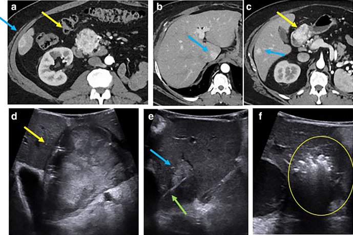
Figure 4.
55-year-old male with metastatic RCC to the pancreas (a, c yellow arrow) and liver (a–c, blue arrows). IOUS image from open approach shows the mass in the pancreas (d, yellow arrow) and hyperechoic caudate liver lesion (e, blue arrow). Patient underwent pancreaticoduodenectomy, wedge resection of hepatic metastatic in segment 4b (not shown) and 6 and intraoperative microwave ablation of hepatic lesions in segment 4a (not shown), caudate, and segments 6/7 for clearance of all metastatic disease. IOUS image demonstrates advancement of a microwave ablation applicator (e, green arrow) into the caudate liver lesion with subsequent ablation and gas cloud with posterior acoustic shadowing encompassing the lesion (f, circle). Due to the small transducer face, it is difficult to lay the needle out along its full length. After this treatment, the patient remains disease free 1 year later. IOUS, intraoperative ultrasound; RCC, renal cellcarcinoma

