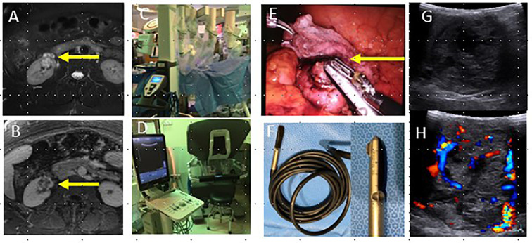
Figure 5.
42-year-old female with complex cystic lesion of the right kidney (arrows, a, b) identified on pre-operative T2W (a) and T1W with contrast (b). Patient underwent robotic partial nephrectomy (c), which utilizes the drop in probe and grasper tool to apply the probe to the lesion (arrow, d) and an ultrasound machine connected to the robot control panel (e). Intraoperative ultrasound images (f) demonstrate a predominantly solid, vascular mass and delineated the proximity of the mass to the renal hilum and major vessels in real time. Surgical pathology demonstrated clear cell RCC. RCC, renalcell carcinoma

