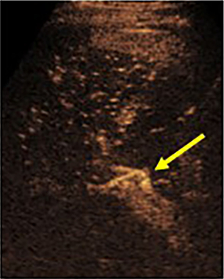
Figure 7.

CEUS in a living donor split liver transplant patient demonstrating a patent hepatic artery (arrow) in the porta that was challenging to see on grayscale ultrasound. The artery was challenging to separate from the adjacent portal vein and CEUS allowed for real-time separation. CEUS, contrast-enhanced ultrasound.
