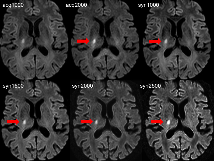
Figure 1.

Acute focal ovaloid ischemic infarct with diffusion abnormality of 9 mm maximal diameter in the posterior limb of the internal capsule along the corticospinal tract on the right side. The lesion is visible on all DW images, however the lesion (red arrows) conspicuity is increased on acq2000, syn1000, syn1500, syn2000 and syn2500 in comparison with the acq1000 image. The subjective image quality is reduced on the acq2000 image and on the syn2500 image.
