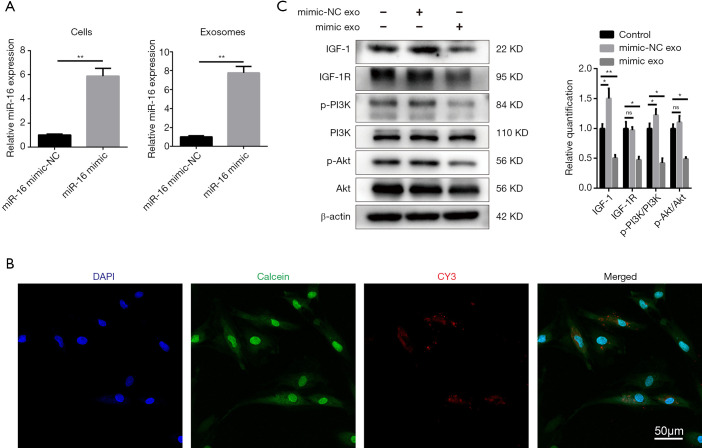
Figure 5.
Exosomes from miR-16 mimic-transfected rat NPC repressed IGF-1 and IGF-1R and the downstream PI3K/Akt pathway in normal rat NPCs. (A) qRT-PCR detection of miR-16 levels from both miR-16 mimic-transfected NPCs and their exosomes. (B) Exosomes from Cy3-labeled miR-16 mimic-transfected NPCs were isolated and incubated with normal NPCs. NPCs incubated with these exosomes exhibited a granular fluorescent pattern within the cytoplasm. Scale bar =50 µm. (C) Western blot showing IGF-1, IGF-1R, and downstream phosphoinositide 3-kinase (PI3K)/protein kinase b (Akt) expression levels. Data are expressed as the mean ± SD, n=3. *P<0.05, **P<0.01 versus the indicated group. Control: NPCs were not treated with anything, mimic-NC exo: exosomes from NPCs transfected with miR-16 mimics-NC, mimic exo: exosomes from NPCs transfected with miR-16 mimics. NPCs, nucleus pulposus cells; qRT-PCR, quantitative real-time polymerase chain reaction; IGF-1, insulin-like growth factor 1; IGF-1R, IGF-1 receptor; SD, standard deviation.

