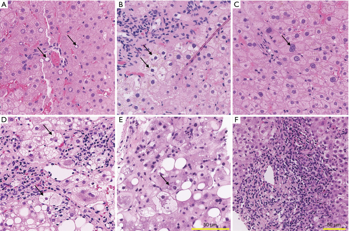
Figure 1.
Liver histology of Wilson disease, non-alcoholic steatohepatitis, alcoholic steatohepatitis, and autoimmune hepatitis. (A-C) Wilson disease. (A) Glycogenated nuclei, adjacent to a small portal tract. (B) Ballooning degeneration. (C) Striking anisonucleosis of hepatocytes. (D) Non-alcoholic steatohepatitis. Numerous ballooned hepatocytes are present adjacent to the central venule, along with lobular inflammation. (E) Alcoholic steatohepatitis. Prominent Mallory-Denk bodies (arrow) are present, as well as discrete foci of lobular injury and cholestasis. (F) Autoimmune hepatitis. Prominent portal inflammation consisting predominantly of lymphocytes and plasma cells, with rare eosinophils and neutrophils. Inflammation extends into the lobule (i.e., interface activity). A rare acidophil body is noted (top right). Sections are stained with hematoxylin and eosin. Scale bar length is 50 µm in all quadrants, one size for quadrants A-E, different size for quadrant F. All images were digitally scanned at 20x magnification.

