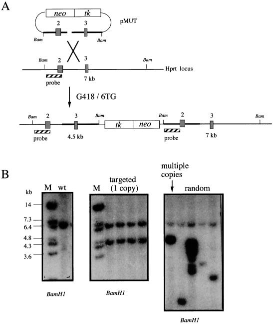
FIG. 3.
Targeting pMUT to the mouse Hprt locus. (A) Schematic diagram of pMUT targeting. Linearization within the Hprt homology region gives rise to an insertion vector. After double selection in G418 and 6TG, a proportion of the clones were targeted with a single copy of pMUT, resulting in the duplication of the target sequences. Hprt exons are represented by numbered boxes. BamHI restriction sites are indicated as Bam. The internal hybridization probe is shown as the striped box. BamHI size fragments are indicated in kilobases. (B) Southern blot analysis of BamHI-digested genomic DNA from wild-type (wt) and G418/6TGr clones by using the internal pMUT probe. Correctly targeted clones containing a single copy of pMUT display a novel band of the predicted size equal in intensity to the wild-type band. Clones displaying hybridization patterns consistent with multicopy and random integration are included for comparison. M, DNA size marker.

