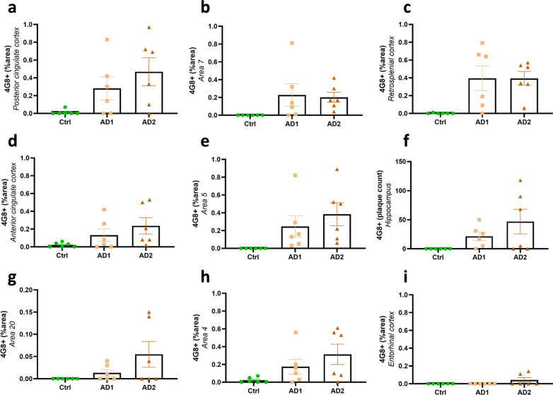
Fig. 3.
Quantification of Aβ deposition following human brain inoculation. Quantification of Aβ load (% 4G8-positive area or Aβ plaque count for the hippocampus) in AD- and Ctrl-inoculated lemurs. Aβ was detected in all of AD-inoculated animals and in none of Ctrl-inoculated lemurs. Some AD-inoculated animals did not display significant amounts of Aβ in some brain regions (e.g. two AD1-inoculated animals did not develop Aβ deposits in the posterior cingulate cortex a), but displayed more Aβ in other regions. Thus, all animals displayed Aβ deposits in at least some regions of their brains. Altogether, animals from both AD-inoculated groups showed Aβ deposition at the inoculation site (posterior cingulate cortex; a) and/or around the needle tract in the parietal cortex (area 7; b). Adjacent regions such as the retrosplenial cortex (c), anterior cingulate cortex (d) and parietal area 5 (e) also displayed Aβ pathology. Aβ plaques were also detected in the hippocampus (f) and other regions more distant from the inoculation sites including the superior temporal cortex (area 20; g) and the frontal cortex (area 4; h). The entorhinal cortex (i) was the only cortical region that did not display Aβ pathology in any groups. No statistical difference was observed between AD1 and AD2-inoculated animals (p > 0.05; Mann–Whitney’s test). Data are shown as mean ± s.e.m

