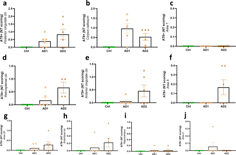
Fig. 9.
Semi-quantitative scoring of neuropil thread pathology following human brain extracts inoculation. Neuropil threads were detected in AD-inoculated animals but not in the Ctrl-inoculated lemurs. AT8-positive neuropil threads were mainly localized at the inoculation sites [posterior cingulate cortex (a) and corpus callosum (b)], but no lesion was observed around the needle tract (area 7; c). They were also induced in juxtaposing regions, such as the retrosplenial cortex (d), anterior cingulate cortex (e), parietal area 5 (f), and in distant regions such as the temporal area 20 (g) and entorhinal cortex (h). Except for one or two animals, neuropil threads were not detected in the hippocampus (i) nor in the frontal cortex (area 4; j). No difference was observed between the two AD-inoculated groups (p > 0.05; Mann–Whitney’s test). Data are shown as mean ± s.e.m

