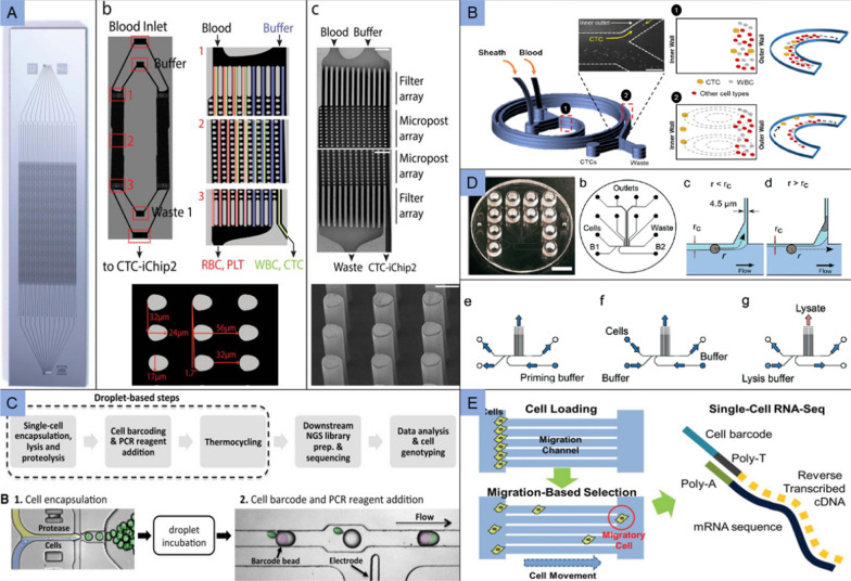
Fig. 6.
Application of microfluidic in cancer diagnosis. A The CTC-iChip composed of two separate and serial chips. Whole blood and buffer inlets enter from top corners, posts deflect nucleated cells away from smaller RBCs, platelets and plasma and toward the buffer. Adapted with permission from Karabacak et al. [134]; B Microfluidics for single cell sorting using DFF. The smaller RBCs and leukocytes exist the outer wall, while the larger CTCs focus along the microchannel inner wall. Adapted with permission from Vaidyanathan et al. [135]; C Protease-based droplet device. Cells are encapsulated with lysis buffer and incubated to promote proteolysis. The droplets containing the cell lysate are paired and merged with droplets containing PCR reagents and barcode-carrying hydrogel beads. Adapted with permission from Pellegrino et al. [141]. D Microfluidic device design and operation. The chip design is based on a hydrodynamic cell trap, and the trapped cell reduces the flow through the trap for the next incoming cell. Adapted with permission from Marie et al. [142]. E Microfluidic chip was performed to isolate migratory cells. Cells are initially positioned at the entrance of migration channels, and loaded cells migrated toward a gradient of serum chemoattractant in the center channel. Adapted with permission from Chen et al. [143]

