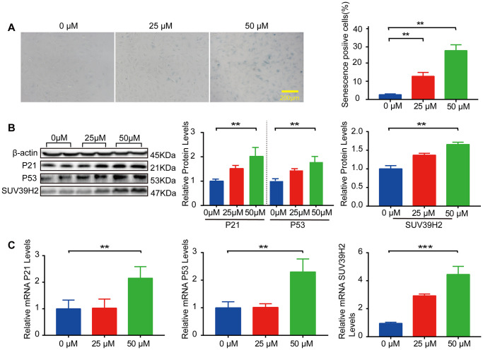
Figure 1.
SUV39H2 is increased in a cardiomyocyte senescence model induced by H2O2. (A–C) Effects of different concentrations of H2O2 on senescence of H9C2 cells. (A) SA-β-Gal staining of H9C2 cells treated with H2O2 concentration in 0, 25, 50 uM groups. (B) Expression of p21, p53, and SUV39H2 in the cells treated with a concentration gradient was detected by western blotting. (C) RT-PCR was used to quantify the expression of p21, p53, and SUV39H2 in above groups. All the experiments have been repeated independently at least 3 times. Scale bars, 200 μm. *P < 0.05, **P < 0.01, ***P < 0.005 when two groups were compared as indicated, or compared to the corresponding control.

