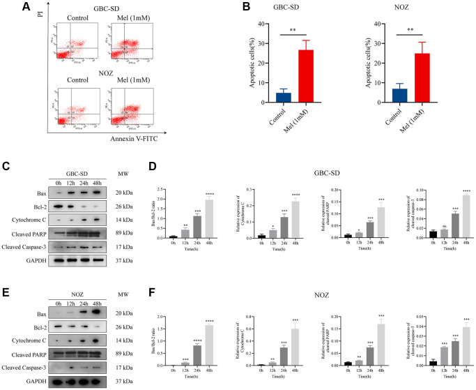
Figure 3.
Melatonin induces apoptosis in gallbladder cancer cells. (A) Apoptosis of GBC-SD and NOZ cells treated with 1 mM melatonin was analyzed by flow cytometry. (B) The percentage of apoptotic cells of GBC-SD and NOZ cells was quantified. (C) Expression of Bax, Bcl-2, cytochrome C, cleaved PRRP, and cleaved caspase-3 was investigated by Western blot after GBC-SD cells were treated with 1 mM melatonin. (D) The relative expression of the apoptotic markers was quantified in GBC-SD cells. (E) Expression of Bax, Bcl-2, cytochrome C, cleaved PRRP and cleaved caspase-3 was investigated by Western blot after NOZ cells were treated with 1 mM melatonin. (F) The relative expression of the apoptotic markers was quantified in NOZ cells. Three biological replicates were performed. Data are presented as mean ± SD. Mel, melatonin; ***P < 0.001; **P < 0.01; *P < 0.05; ns, no significance.

