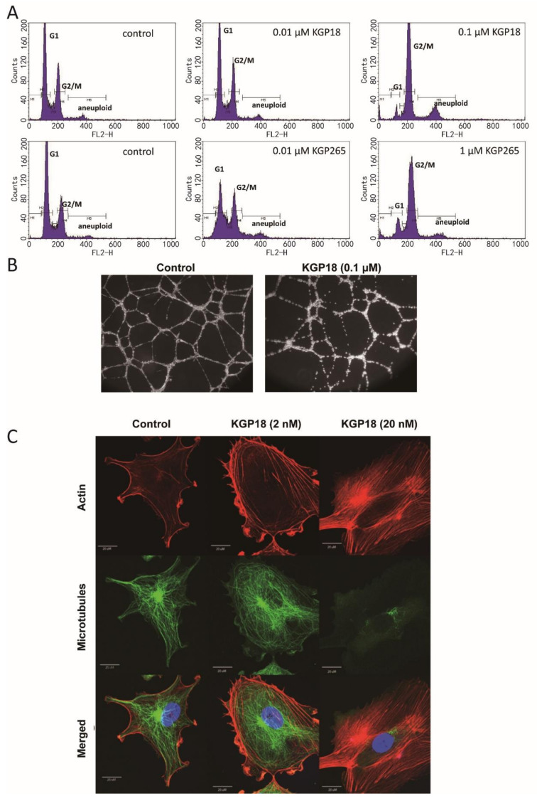
Figure 2.
KGP18 treatment of MDA-MB-231 cells and activated HUVECs. (A) KGP18 (active agent) and the corresponding phosphate prodrug KGP265 induced concentration-dependent G2/M arrest in MDA-MB-231 cells, as assessed by flow cytometry. (B) KGP18 treatment disrupted capillary-like endothelial networks pre-established with HUVECs on Matrigel. (C) Monolayers of rapidly growing HUVECs underwent concentration-dependent changes in the cell morphology with KGP18 treatment (2 h) that demonstrated a loss of the microtubule structure and increased the bundling of filamentous actin into stress fibers. Representative confocal images of endothelial cells stained for α-tubulin (FITC), actin (Texas Red) and the nuclei (DAPI). Bar: 20 µm. HUVEC, human umbilical vein endothelial cell.

