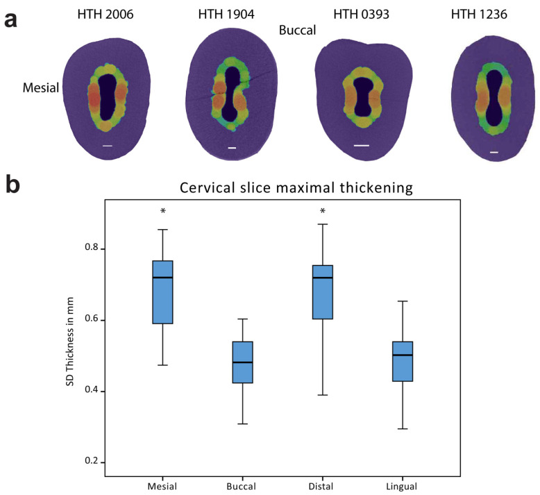
Figure 3.
Mesio-distal thickening of SD. (a) Horizontal slices at −1 mm below the CEJ from the four representative teeth. Buccal and mesial directions are on the top and on the right, respectively. SD thickness values are based on the thickness legend. (b) Box plot of the SD thickness 1 mm below the CEJ at mesial, buccal, distal, and lingual directions. n =30. * Indicating significant difference at p < 0.05. Significance was found between the mesial and distal to buccal and lingual.

