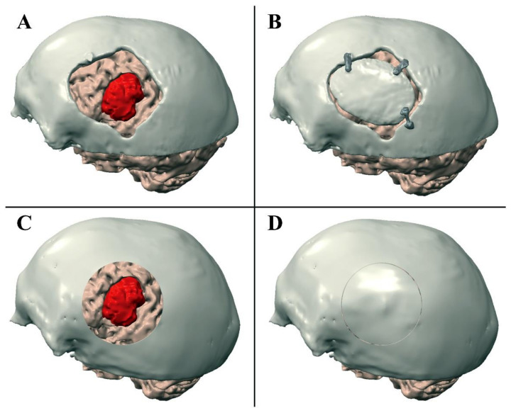
Figure 3.
Performed craniotomy (A,B) and VOSTARS planning (C,D). (A) Shows the post-operative skull model was registered to the preoperative brain model. (B) Shows the position of the bone flap cranioplasty with polymethylmethacrylate. (C) Shows the craniotomy planned with VOSTARS. (D) Shows the result of the ideal post-operative bone flap cranioplasty on the manikin’s head.

