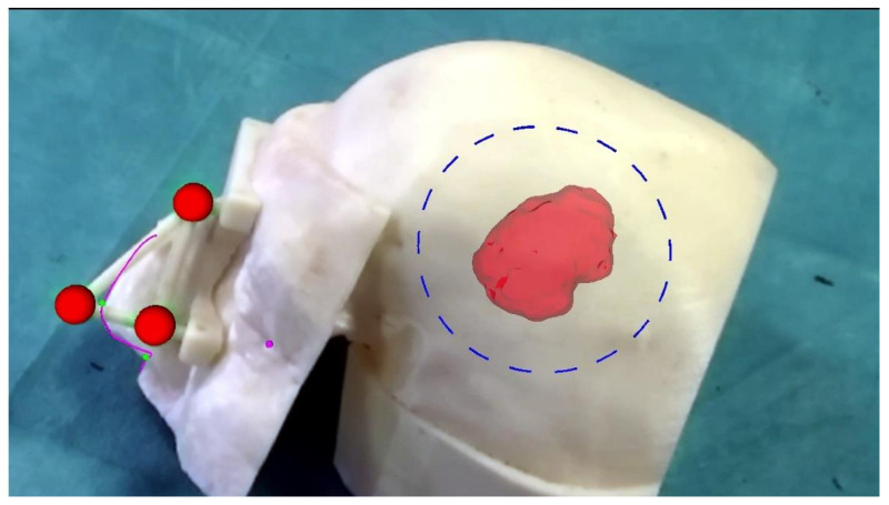
Figure 6.
Augmented reality scene with 3D reconstruction of the meningioma (the semi-transparent red structure), the planned craniotomy (the blue dashed line), the optical markers (the red spheres), the reference landmarks (the small pink and green spheres) and the nose profile (the pink line). The last two virtual elements are used as reference landmarks to verify the accuracy of the image-to-patient registration. The ideal pre-operative and intra-operative procedural steps are outlined in Figure 7.

