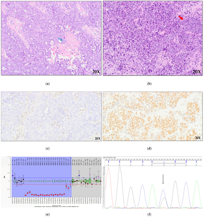
Figure 1.
Example of two representative poorly differentiated sinonasal carcinomas. On the left side, an NKSCC ((a), haematoxylin-eosin stain, x20) with ribbon-like growth pattern, absent maturation and necrotic areas (blue arrows), showing total loss of SMARCB1 protein in the tumor area except for the normal stromal cells (c) and biallelic loss of SMARCB1 gene ((e), red dots). On the right side, a LCNEC (b) composed by medium-large cells arranged in organoid nests (red arrow), showing the presence of SMARCB1/INI1 protein (d) with intense nuclear positivity (brown stain) and IDH2 p.Arg172Thr mutation ((f), black arrow).

