Figure 1.
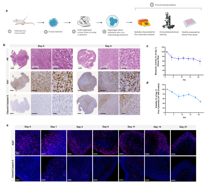
Organotypic culture. (a) Project workflow. Mice are injected with MosJ osteosarcoma cells; when desired mass is reached, mice are sacrificed, and the tumour is resected. Tumour slices are cut using a vibratome and cultured with/without chemotherapy treatments. Organotypic slices are fixed for immunohistochemical staining, dissociated for flow cytometry analysis, or assessed by Alamar blue assay. (b) 5 μm slices of fixed paraffin embedded organotypic sections from day 0 and day 4 are stained with H&E, Ki67, and cleaved caspase-3. (c) Metabolic activity and (d) cell viability of organotypic slices were measured for 21 days (Day 1, 4, 8, 11, 14, 17 and 21) and normalised to day 1; data is expressed as Mean ± SD of n = 6 samples and n = 3 samples, respectively. (e) Fluorescent microscopy on 5 μm slices of fixed paraffin embedded organotypic sections from day 0 to day 21. Samples are labelled with Ki67 (cell proliferation, pink) or cleaved caspase-3 (apoptosis, pink), and DAPI (nuclei, blue).

