Figure 1.
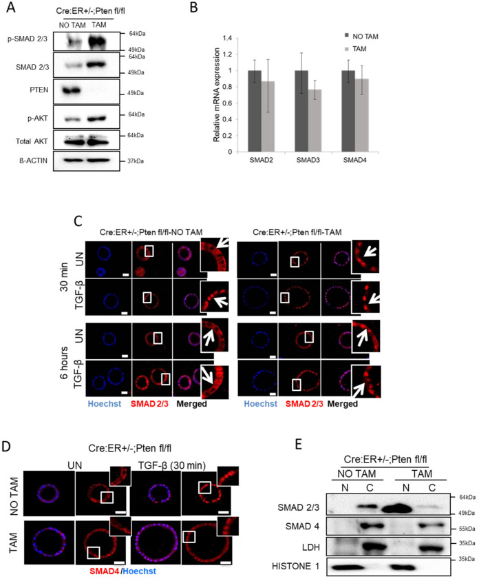
PTEN deficiency triggers constitutive nuclear localization of SMAD2/3 in endometrial organoids. (A) Western blot analysis of p-SMAD2/3, SMAD2/3 and p-AKT on lysates from Cre:ER+/−;PTEN fl/fl organoid cultures treated (TAM) or not (NO TAM) with tamoxifen. Membranes were also blotted with PTEN antibody to show its total deletion after tamoxifen treatment. Membranes were also reblotted with total AKT and β-actin antibodies to show equal protein loading. A representative image of n = 3 biological replicates is shown. (B) RT-qPCR analysis of SMAD2, SMAD3, SMAD4 mRNA corresponding to organoid cultures from Cre:ER+/−;PTENfl/fl organoid cultures treated (TAM) or not (NO TAM) with tamoxifen. (C) SMAD2/3 immunofluorescence on Cre:ER+/−;PTENfl/fl organoid endometrial organoids incubated (TAM) or not (NO TAM) with tamoxifen delete PTEN and then treated for 30 min with 10 ng/mL TGF-β, 6 h or left untreated (UN). Organoid cultures were counterstained with Hoechst to show nuclei. Magnification images of framed regions of sample are shown to show SMAD2/3 cellular localization. Arrows indicate presence or absence of nuclear staining. Scale bars: 25 μm. Data are from n = 3 experimental replicates (independent organoid cultures). (D) Representative SMAD4 immunofluorescence on Cre:ER+/−;PTENfl/fl endometrial cultures incubated (TAM) or not (NO TAM) with tamoxifen to induce PTEN deletion and then treated with 10 ng/mL TGF-β for 30 min or untreated (UN). Magnification images of framed regions of the samples show SMAD4 cellular localization. Data are from n = 3 experimental replicates (independent organoid cultures). Scale bars: 25 μm. (E) Western blot analysis of SMAD2/3 and SMAD4 on nuclear (N) and cytosolic (C) fractions of Cre:ER+/−;PTENfl/fl 3D cultures treated (TAM) or not (NO TAM) with tamoxifen. Membranes were also probed with Histone H1 and LDH to demonstrate correct nuclear and cytosolic fractionation. A representative image of n = 3 biological replicates is shown. Original Western blot data is shown in Supplementary Materials.

