Figure 2.
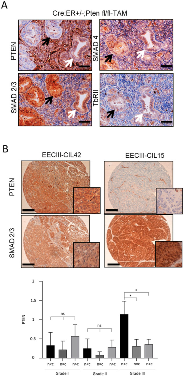
PTEN deficiency leads to nuclear constitutive localization of SMAD2/3 in vivo. (A) PTEN and SMAD2/3 immunohistochemistry on formalin-fixed paraffin-embedded endometrial tissue sections from 12-week-old Cre:ER+/−;PTENfl/fl mice that have been injected with tamoxifen (TAM) to induce PTEN deletion (4 weeks). 20× images. Black arrows indicate PTEN-negative glands that display nuclear SMAD2/3 staining. White arrows indicate glands that keep PTEN expression and display a more cytoplasmatic SMAD2/3 staining. Scale bar: 100 μm. (B) Representative images of PTEN and SMAD2/3 immunohistochemistry on formalin-fixed paraffin-embedded endometrial tissue sections from two human grade III endometrial carcinomas. Correlation of PTEN expression values and SMAD2/3 expression in the nucleus in grade I, grade II and grade III EECs. (n < c) designates more expression in the cytoplasm than in the nucleus, (n = c) equal expression and (n > c) higher expression in the nucleus than in the cytoplasm (bottom graph). Vertical bars represent ± standard error. Plot evidences a reduction of PTEN expression when the expression of SMAD2/3 in the nucleus is higher than in the cytoplasm (p = 0.02) in n = 37 EEC grade III (20× images and magnifications). Scale bar: 100 μm. * p < 0.05 by t-test analysis.

