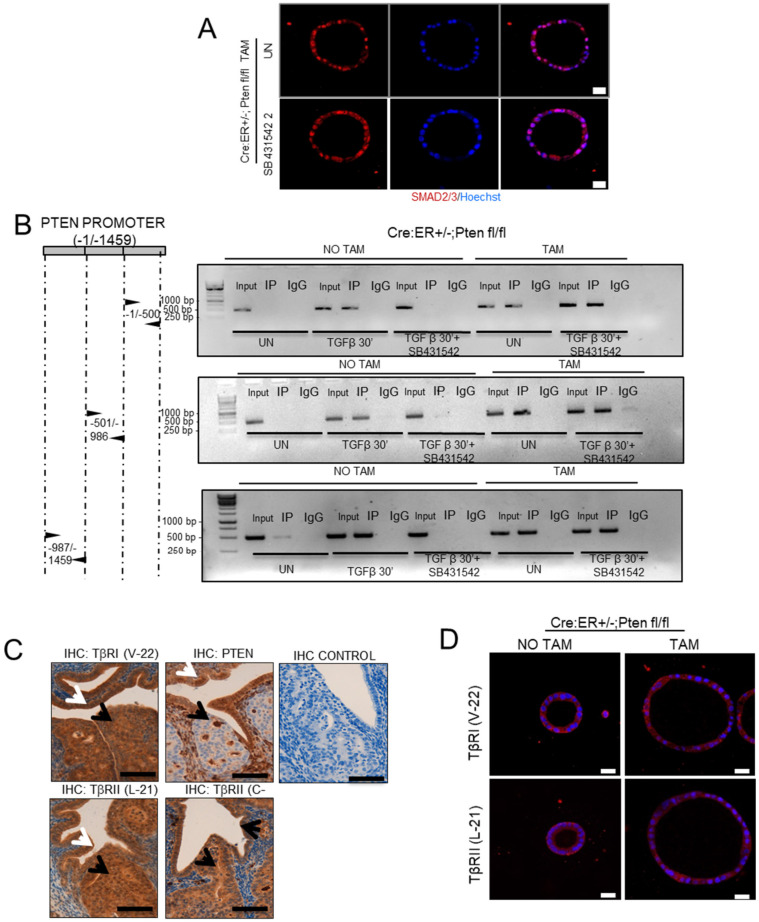
Figure 3.
SMAD2/3 nuclear translocation is independent of TGF-β receptor. (A) SMAD2/3 immunofluorescence on Cre:ER+/−;PTENfl/fl organoids incubated with tamoxifen (TAM) to induce the deletion of PTEN and treated with SB431542 10 µM or left untreated (UN). To evidence morphology, nuclei were counterstained with Hoechst. Scale bars: 25 μm. Data are from n = 3 experimental replicates (independent organoids cultures). (B) ChIP of SMAD2/3 binding to PTEN promoter. Organoids from Cre:ER+/−;PTENfl/fl treated with tamoxifen (TAM) or not (NO TAM) to delete PTEN were pretreated or not with SB431542 10 µM for 2 h and then stimulated for 30 min with TGF-β. Untreated (UN) or TGF-β stimulated organoids were lysed and immunoprecipitated (IP) with control antibody (IgG) or SMAD2/3. Immunoprecipitates were subjected to PCR analysis using primers to amplify three ~500 bp segments of PTEN promoter (−1/−500, −501/−986, −987/−1459). (C) TbRI (antibody V-22) and TGFβRII (two different antibodies L-21 and C-16), PTEN immunohistochemistry and negative control on serial sections of endometrial tissue were obtained from 1-week-old Cre:ER+/−;PTENfl/fl mice; injected with tamoxifen to induce PTEN deletion. Mice were sacrificed 4 weeks after tamoxifen injection. Black arrows indicate PTEN-deficient glands in serial sections. White arrows indicate PTEN-positive gland in serial sections. (40× magnifications; n = 10 Cre:ER+/−;PTENfl/fll-TAM mice). Scale bar: 100 µM. (D) TGFβRI (antibody V-22, red) and TGFβRII (antibody L-21, red) immunofluorescence analysis on Cre:ER+/−;PTENfl/fl organoids incubated (TAM) or not (NO TAM) with tamoxifen to induce PTEN deletion. Organoids were counterstained with Hoechst (Blue) to evidence nuclei. Scale bars: 25 μm. Data are from n = 3 experimental replicates (independent organoid cultures).

