Figure 1.
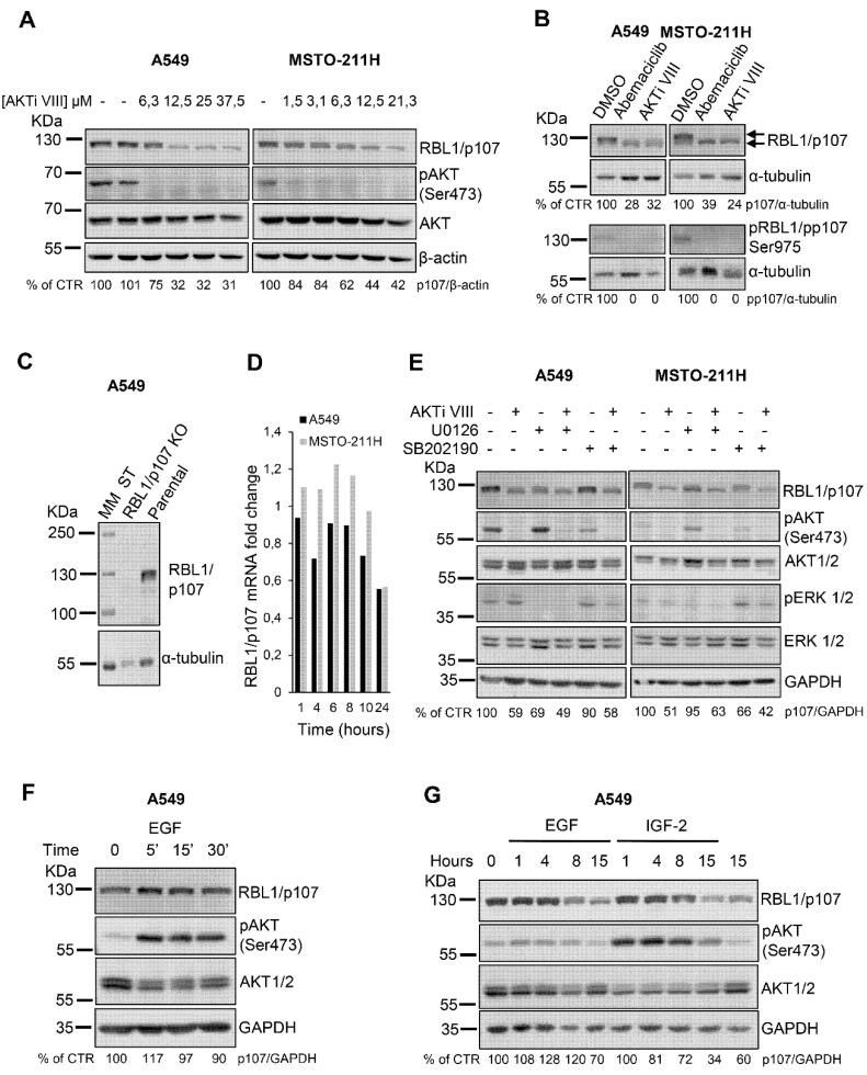
The AKT inhibitor AKTi VIII reduces RBL1/p107 levels in A549 and MSTO-211H cells. (A). A549 and MSTO-211H cells were treated with the indicated concentrations of the AKT inhibitor AKTi VIII for 16 h. RBL1/p107 levels were analyzed in cell lysates by immunoblot. (B). A549 and MSTO-211H cells were treated with 0.2 µM Abemaciclib or 12.5 µM AKTi VIII for 16 h. Levels of total and phosphorylated p107 (ser975) were analyzed by immunoblot. Arrows indicate the two major bands corresponding to hyper- and hypo-phosphorylated RBL1/p107. (C). RBL1/p107 levels were analyzed in cell lysates of parental and A549 cells with RBL1/p107 knock-out by CRISPR/Cas9. MM: molecular mass. (D). RBL1/p107 mRNA levels were determined by RT-PCR in A549 and MSTO-211H cells treated with 12.5 µM AKTi VIII for the indicated time points. (E). RBL1/p107 levels were determined by immunoblot in A549 and MSTO-211H cells preincubated with the MEK1/2 inhibitor U0126 or the p38 inhibitor SB202190, at the concentration of 10 and 20 µM, respectively, for 2 h and then treated with the same concentrations of U0126 or SB202190 alone or in combination with 12.5 µM AKTi VIII for 16 h (F). A549 cells were treated with 50 ng/mL EGF for the indicated time and RBL1/p107 protein levels analyzed by immunoblot. (G). RBL1/p107 levels were analyzed in cell lysates of A549 cells treated for the indicated time with 50 ng/mL EGF or 10 nM IGF-2. Western blot quantification was performed using the ImageJ program, normalized to β-actin, α-tubulin or GAPDH expression and reported as % of control (CTR). All experiments were performed three times.

