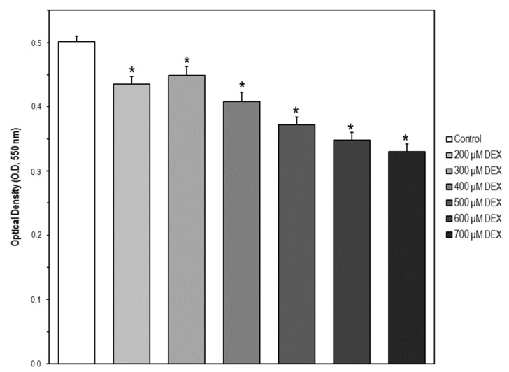
Figure 5.
Cell model of apoptosis by MC3T3-E1 cell exposure to dexamethasone (DEX). Cells were treated with DEX in a concentration range of 0–700 μM for 24 h. Then, cell viability was measured by the MTT assay. The data are expressed as the mean ± S.E.M. of three independent experiments. Significant differences were found in optical density (OD, 550 nm) values elicited by every DEX concentration compared to the control untreated cells (Newman–Keuls test, * p < 0.05 vs. control).

