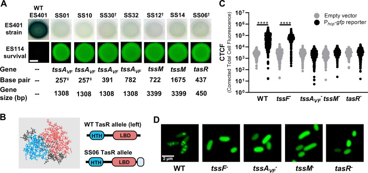
FIG 2.
Isolated transposon mutants are unable to activate T6SS transcriptional reporters or build sheaths. (A) Qualitative reporter activity for Phcp-lacZ in wild-type ES401 and transposon mutants. “Gene” and “Base pair” indicate the location of transposon insertion for each mutant based on the ES401 reference genome (52). Daggers indicate representative mutants from tssAVF, tssM, and tasR that were selected for further characterization. Shown are fluorescence microscopy images of ES114 at 24 h following coincubation with each mutant. The scale bar is 2 mm. Images were taken after 24 h on LBS–X-Gal (reporter) or LBS (fluorescence). Assays were performed at least three times (n = 4), and a representative experiment is shown. (B) Predicted structure of TasR using Quark (80, 81). Shown are the N-terminal helix-turn-helix domain (blue), C-terminal ligand binding domain (pink), and C-terminal extension in the SS06 mutant (CLLYTSAAALGLAVVLQGP [gray]). (C) Quantitative single-cell reporter activity for Phcp-gfp in wild-type ES401, a structural tssF mutant, and representative regulatory mutants. Cells carrying plasmid pAS2028 were imaged immediately following a 5-h incubation on LBS agar, and the corrected total cell fluorescence (CTCF) was calculated for single cells. The assay was performed two times (n > 1,000 cells/treatment), and all data are shown. Circles indicate CTCF measurements from single cells. *, P < 0.0001 (one-way analysis of variance [ANOVA] followed by a Tukey’s multiple-comparison test comparing the empty vector to the Phcp-gfp reporter in each strain). (D) Representative green fluorescence images of the ES401 wild-type, a structural tssF mutant, and representative regulatory mutant cells carrying the TssB_2-GFP expression vector (pSNS119) after incubation on LBS agar for 5 h. LBS medium was supplemented with 0.5 mM IPTG (isopropyl-β-d-thiogalactopyranoside) to induce expression of TssB_2_GFP.

