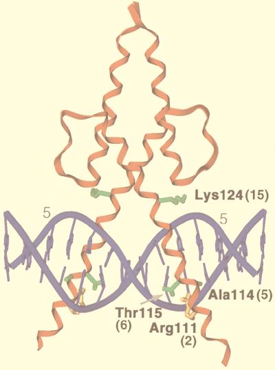
FIG. 1.
A MyoD-DNA complex. In this X-ray crystallographic structure (35), a MyoD homodimer is bound to the sequence AACAGCTGTT, which corresponds to its preferred recognition consensus (9). Residues are numbered as in full-length MyoD, and their positions as specified in Fig. 2 and the text are indicated in parentheses. Binding site positions ±5 (numbered as in Fig. 3A) are indicated by grey numerals. Side chains are shown only for the myogenic residues (green) (18) and Arg 111 (R2) (gold).

