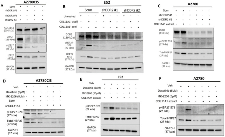
Figure 2.
COL11A1 upregulates HSP27 phosphorylation and expression through activation of DDR2/integrin α1β1-Src-Akt signaling in ovarian cancer. (A) Western blot of the phosphorylated (at S78) and total HSP27, DDR2, and COL11A1 in A2780CIS-scrambled (scrm) and A2780CIS-shDDR2 cells. (B) Western blot of the phosphorylated (at S78) and total HSP27 in ES2-scrm and ES2-shDDR2 #1 and #2 on COL11A1-positive and -negative A204-derived decellularized matrices and control conditions. (C) Western blot of the phosphorylated (at S78) and total HSP27 in A2780-scrm and A2780-shDDR2 in the COL11A1-positive extract and control conditions. (D) A2780CIS-scrm and A2780CIS-shCOL11A1 cells treated with Dasatinib (5 µM) and MK-2206 (5 µM) for 72 h. (E) ES2 cells cultured on the COL11A1-positive extract and control conditions treated with Dasatinib (5 µM) and MK-2206 (10 µM) for 72 h. (F) A2780 cells cultured on the COL11A1-positive extract and control conditions treated with Dasatinib (5 µM) and MK-2206 (5 µM) for 72 h. GAPDH was used as a loading control for the Western blots. All experiments were performed in the standard experiment conditions (cells cultured in the above conditions for 3 days in 1% FBS medium after overnight serum starvation). The uncropped Western blot figures are presented in Figure S6.

