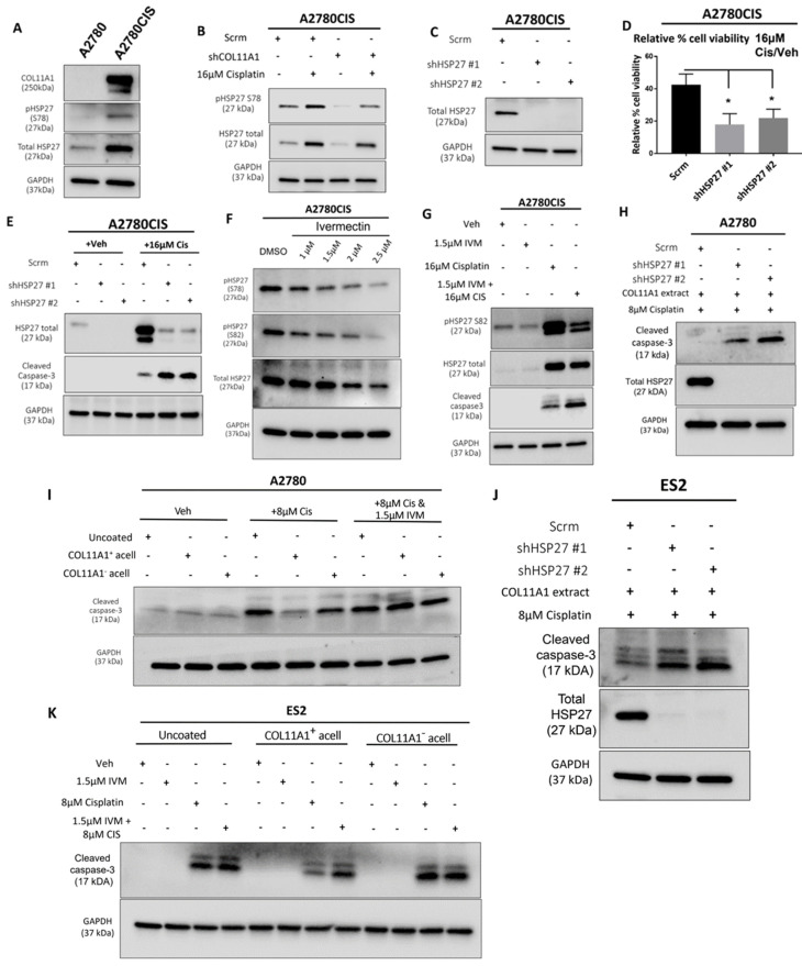
Figure 3.
HSP27 mediates COL11A1-induced cisplatin resistance in ovarian cancer cells. (A) Western blot of the phosphorylated (at S78) and total HSP27 in A2780 and A2780CIS. (B) Western blot of the phosphorylated (at S78) and total HSP27 in A2780CIS-scrm and A2780CIS-shCOL11A1 treated with 16 µM cisplatin. (C) Western blot of the total HSP27 in A2780CIS-scrm and A2780CIS-shHSP27 #1 and #2. (D) Relative cell viability (measured by Cell titer glo) of A2780CIS-scrm and A2780CIS-shHSP27 #1 and #2 treated with 16 µM cisplatin compared with the respective vehicle-treated control groups (n = 3). (E) Western blot of total HSP27 and cleaved caspase-3 in A2780CIS-scrm and A2780CIS-shHSP27 #1 and #2 and treated with the vehicle treatment or 16 µM cisplatin. (F) Western blot of the phosphorylated (at S78 and S82) and total HSP27 in the vehicle and 1 µM, 1.5 µM, 2 µM, and 2.5 µM ivermectin (IVM)-treated A2780CIS cells. (G) Western blot of the phosphorylated (at S82) and total HSP27 and cleaved caspase-3 in the vehicle, 1.5 µM ivermectin (IVM), 16 µM cisplatin, and combination-treated (1.5 µM ivermectin and 16 µM cisplatin) A2780CIS cells. (H) Western blot of the total HSP27 and cleaved caspase-3 in A2780-scrm and A2780-shHSP27 #1 and #2 cultured on a COL11A1 extract and treated with 8 µM cisplatin. (I) Western blot of and cleaved caspase-3 in A2780 cells cultured on COL11A1-positive and -negative A204-derived decellularized matrices and control conditions treated with 8 µM cisplatin (Cis) and the combination (1.5 µM ivermectin and 8 µM cisplatin (IVM + Cis)) and vehicle treatments. (J) Western blot of the total HSP27 and cleaved caspase-3 in ES2-scrm and ES2-shHSP27 #1 and #2 cultured on a COL11A1 extract and treated with 8 µM cisplatin. (K) Western blot of cleaved caspase-3 in ES2 cells cultured on COL11A1-positive and -negative A204-derived decellularized matrices and control conditions treated with 1.5 µM ivermectin (IVM), 8 µM cisplatin (Cis), and the combination (1.5 µM ivermectin and 8 µM cisplatin (IVM + Cis)) and vehicle treatments. GAPDH was used as a loading control for the Western blots. All experiments were performed in the standard experiment conditions (cells cultured in the above conditions for 3 days in 1% FBS medium after overnight serum starvation). Error bars indicate the standard deviation. *, p value < 0.05. The uncropped Western blot figures are presented in Figure S7.

