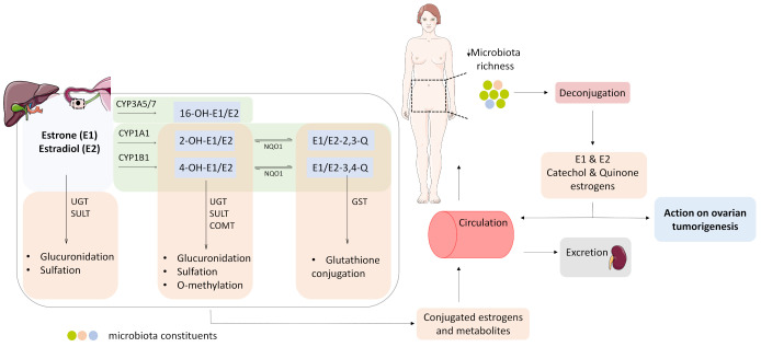
Figure 2.
Estrogen metabolism in the liver and in the ovarian tumor tissue. Oxidation (light green boxes) is catalyzed by cytochrome P450 enzymes, and gives rise to catechol-estrogens: 2- and 4- OH estrone/estradiol, and 16-OH-estrone/estradiol (2-OH-E1/E2 and 4-OH-E1/E2, 16-OH-E1/E2). 2- and 4- catechol-estrogens are further oxidized to DNA-damaging quinones (E1/E2-2,3-Q and E1/E2-3,4-Q). Conjugation (light orange boxes) involves glucuronidation, catalyzed by UDP glucuronosyl transferases (UGT2B7), sulfation, catalyzed by sulfotransferases (SULT1A1, SULT1E1, SULT2B1), methylation, catalyzed by catechol-O-methyl transferase (COMT), and glutathione conjugation, catalyzed by glutathione S-transferases (GSTP1). The conjugated estrogens and their metabolites enter the systemic circulation and are eventually excreted from the body through urine and feces. The microbiome, more precisely the estrobolome, actively modulates the estrogen levels in the body. The microbiome change observed in OC patients, might contribute to the pool of estrogens available to the estrogen-responsive ovarian tumor by de-conjugating already conjugated estrogens and estrogen metabolites, hence to ovarian tumorigenesis. COMT, catechol-ortho-methyl transferase; CYP1A1, cytochrome P450 1A1; CYP1B1, cytochrome P450 1B1; CYP3A5, cytochrome P450 3A5; CYP3A7, cytochrome P450 3A7; GST, glutathione-s-transferase; NQO1, NAD(P)H quinone oxidoreductase 1; SULT, sulfotransferase; UGT, uridine 5’ diphospho-glucuronosyl transferase. Parts of the figure were drawn by using pictures from Servier Medical Art. Servier Medical Art by Servier is licensed under a Creative Commons Attribution 3.0 Unported License (https://creativecommons.org/licenses/by/3.0/) (accessed on 7 May 2021).

