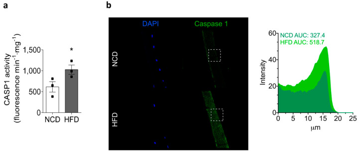
Figure 5.
HFD increases caspase-1 activity in the skeletal muscle. Gastrocnemius muscles were collected from NCD- or HFD-fed mice, homogenized, and caspase-1 activity was measured. Values represent the mean ± SEM (n = 3). * p < 0.05, determined by Student’s t-test. (a) Caspase-1 activity in muscle lysates was measured with a caspase-1 fluorometric kit. (b) Representative confocal images of isolated adult FDB fibers from NCD- and HFD-fed mice illustrate the localization of caspase-1 (green). Nuclei were stained with DAPI (blue). The area under the curve (AUC) was measured on white squares, and represented as fluorescence intensity. Scale bar: 20 μm.

