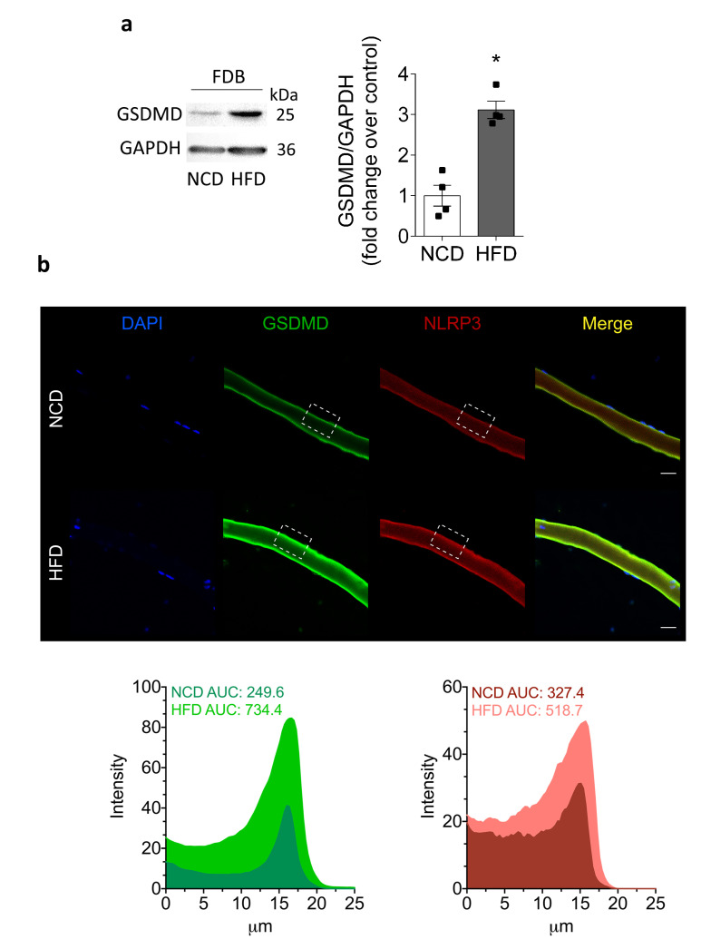
Figure 6.
Cleaved GSDMD increases in HFD-fed mice. We dissected and homogenized FDB muscles from NCD- or HFD-fed mice and assessed the GSDMD level by Western blot. We also studied GSDMD subcellular location in fixed isolated fibers by immunofluorescence. (a) Representative Western blot and quantification showing cleaved GSDMD; 60 µg of lysate was loaded in each lane. The GSDMD bands were observed at 25 kDa. Values represent the mean ± SEM (n = 4). * p < 0.05, determined by Student’s t-test. (b) Representative confocal images of isolated adult FDB fibers from NCD- and HFD-fed mice illustrating the localization of GSDMD (green) and NLRP3 (red). DAPI staining of the nuclei is shown in blue. The area under the curve (AUC) was measured on white squares, and represented as fluorescence intensity. Scale bar: 20 μm.

