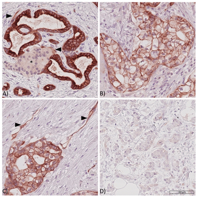
Figure 1.
Expression of the insulin receptor in PDAC tissues. Representative PDAC tissue samples showing (A) high cytoplasmic (cCC-IR 2+), high membranous (mCC-IR 2+) and low vascular (arrow heads, VIR 1+) insulin receptor expression with pancreatic cancer cells surrounding a pancreatic islet (asterisk*), (B) low cytoplasmatic (cCC-IR 1+) and low (mCC-IR 1+) as well as high membranous (mCC-IR 2+) insulin receptor expression, (C) low (mCC-IR 1+) as well as high (mCC-IR 2+) membranous, weak cytoplasmic (cCC-IR 1+) and strong vascular (VIR 2+, arrowheads) insulin receptor expression and (D) absent tumoral or vascular insulin receptor expression. Anti-insulin receptor immunostaining, hematoxylin counterstaining. Original magnification A-D: 400×.

