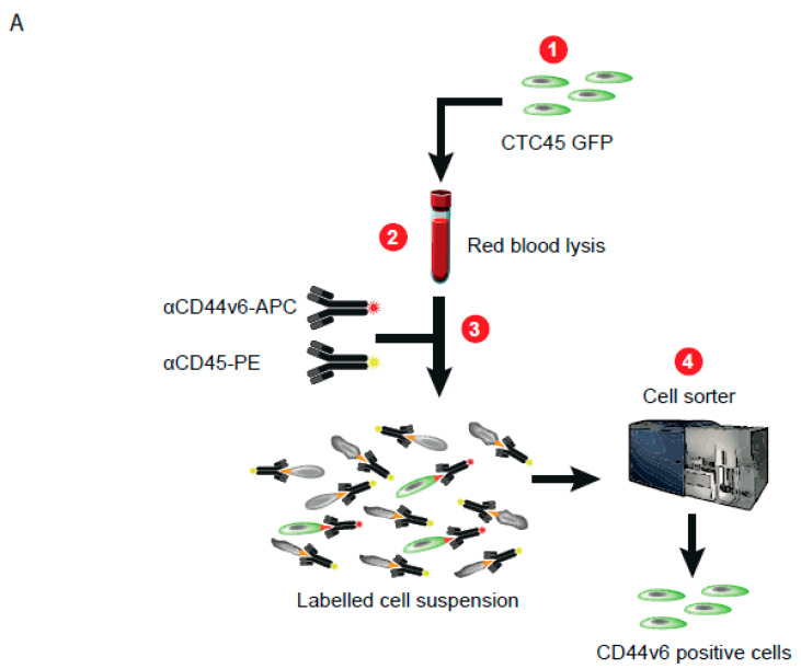
Figure 3.
Cytometry-based isolation following CD44v6 staining efficiently discriminated tumor cells from hematopoietic cells. (A) Flowchart summarizing the strategy used to purify CTC in patient blood samples through fluorescent-activating cell sorting (FACS). Detailed protocol is described in the Materials and Methods section. (B). Photos of GFP-positive CTCs cultured after cell sorting following staining with an antibody directed against CD44v6, observed with a microscope (fluorescent for the left photo and bright field for the middle one; the right photo is merged). Scale bars represent 50 µm.


