Figure 4.
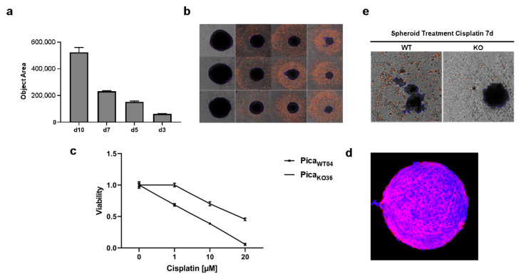
Absence of VRAC expression protects 3D tumor spheroids against cisplatin-induced cancer cell death. (a,b) Automated high content microscopy to visualize tumor spheroid growth. (a) Mean object sizes of spheroids (n = 8) automatically determined by high-content screening microscope Array Scan VTI. (b) Exemplary images of automatically detected regions of interest (ROI). Magnification, 5-fold. (c) Application of spheroids to confirm VRAC’s relevance for cisplatin resistance even in tumor mimicking 3D cultures. Spheroids were treated for 72 h, and viability was normalized to untreated controls. (d) Microarchitecture of spheroids visualized by deep-tissue two photon excitation (2PE) microscopy. EpCAM was detected by specific fluorescent antibodies (red), nuclei were stained with Hoechst dye (blue). Magnification, 20-fold. (e) Microscopy demonstrates that PicaWT04 (WT) spheroids were killed and disassemble under cisplatin treatment compared to PicaKO36 (KO) spheroids. Magnification, 5-fold.

