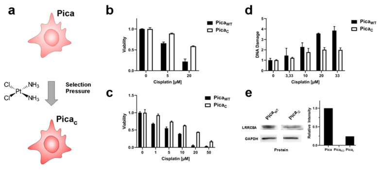
Figure 5.
VRAC levels affect cisplatin sensitivity also in naturally occurring cisplatin-selected cells. (a) Scheme to illustrate the establishment of cisplatin-resistant PicaC cells by chronic exposure to cisplatin. (b,c) 2D/3D PicaC cell models to demonstrate cisplatin resistance. Cells (b) or spheroids (c) were drug treated (cells—48 h; spheroids—72 h), and viability was normalized to untreated controls. (d) Resistant PicaC cells show a lower number of cisplatin-induced DNA damage events (γH2AX foci) per cell as automatically quantified by high-throughput microscopy. γH2AX foci stained by specific fluorescent antibodies. (e) Immunoblot analysis confers downregulation of VRAC (LRRC8A) protein levels in resistant PicaC cells. GAPDH served as loading control. Data from one representative experiment of three independent experiments shown. For immunoblot analysis of PicaKO36 cells, refer to Figure 2b. For uncropped blots, refer to Supplementary Materials.

