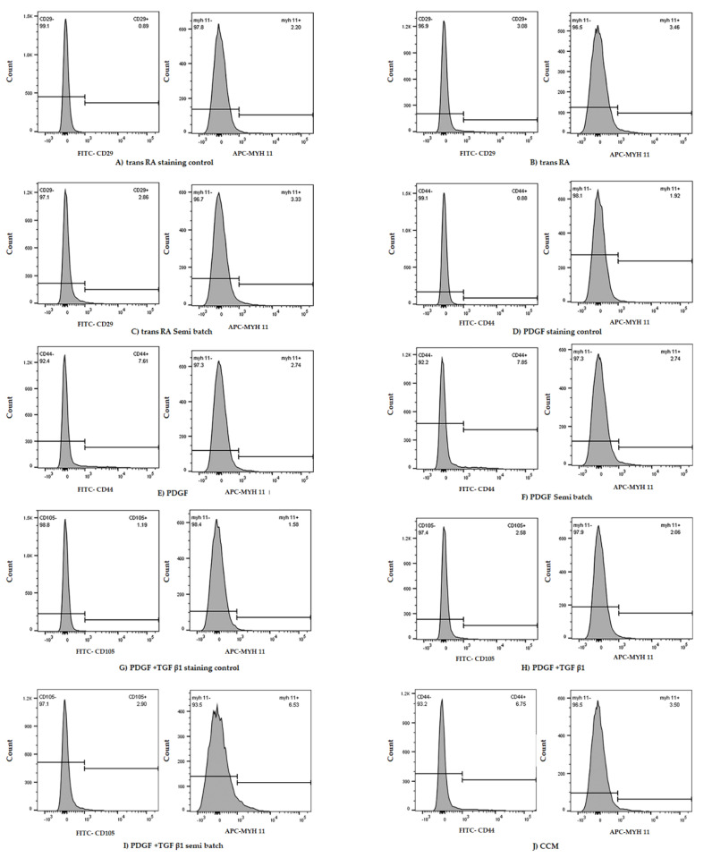
Figure 3.
Representative flow cytometry data from M1 cell line subjected to 7 days in different DM treatments (A–J). Staining control samples were subjected to only secondary antibodies (A,D,G). Each non-control tube was stained with two primary antibodies which denoted SMC (MYH II) and MSC (CD29, CD44, CD105) markers, respectively (B,C,E,F,H,I). The cell line cultured in cell culture medium (CCM) is considered an experimental control. In this trial, the samples subjected to PDGF and TGF β1 (semi-batch) (I) elicited the highest MYH11 expression.

