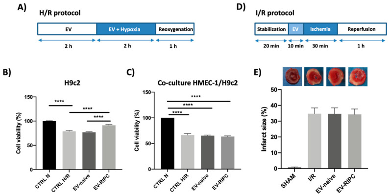
Figure 3.
Impact of EVs in vitro and ex vivo. (A) Timeline of in vitro H/R protocol. H9c2 or co-culture of H9c2 and HMEC-1 were subjected to 2 h of hypoxia, followed by 1 h of reoxygenation. EVs were infused for 2 h before hypoxia and during hypoxia. (B) Cell viability on H9c2 cells subjected to H/R protocol, after treatment with EV-naive or EV-RIPC; data were normalized to the mean value of normoxic control. (**** p < 0.0001 CTRL N vs. CTRL H/R; **** p < 0.0001 CTRL H/R vs. EV-RIPC; **** p < 0.0001 EV-naive vs. EV-RIPC). (C) Cell viability on H9c2 co-cultured with HMEC-1 in trans-well assay; data were normalized to the mean value of normoxic control. (D) Timeline of ex vivo I/R protocol. The hearts were subjected to 30 min of global, normothermic ischemia, followed by 60 min of reperfusion. EVs were infused for 10 min before ischemia. (E) Infarct size in isolated rat hearts treated as indicated. The necrotic mass was measured at the end of reperfusion and reported as percentage of the left ventricle mass (LV; % IS/LV).

