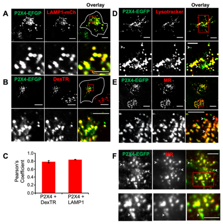
Figure 1.

Colocalization of P2X4 receptors with endolysosomal markers. rP2X4-EGFP receptors were expressed in NRK cells, and live cells were imaged by confocal fluorescence microscopy 48 h post transfection after co-labeling lysosomes with a variety of different markers. These include (A) LAMP1-mCherry (LAMP1-mCh), which was co-transfected with rP2X4-EGFP, and (B) Texas Red-labeled Dextran (DexTR) incubated for 5 h and chased for 2 h at 37 °C. (C) Calculation of Pearson’s coefficient showed the high degree of colocalization between rP2X4 and LAMP1-mCh/DexTR. Results are the mean ± SEM from three independent experiments, total number of cells, n = 40. (D) Cells were incubated with Lysotracker (50 nM) for 10 min at 37 °C and, in (E,F), with a fluorescent substrate of cathepsin B, Magic RedTM (MR) for 5 min. Cells in (F) were imaged using TIRF microscopy to show only those rP2X4- and MR-positive compartments located within ~100 nm of the plasma membrane. Arrowheads indicate examples of compartments that are labeled only by rP2X4 or MR. Scale bars are 10 µm (5 µm for enlargements).
