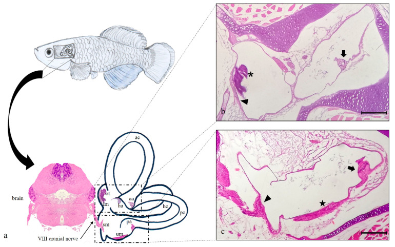
Figure 1.
(a) graphical representation of N. guentheri inner ear that includes the three semicircular canals: anterior (ac), horizontal (hc), and posterior (pc) with their respective crista ampullaris (aa, ha, pa). Furthermore, the horizontal canal contains the macula of lagena (ml) and otholith (ot), while the posterior canal contains saccular macula (sm) and utricular macula (um). The brain from which branches off the VIII° cranial nerve that innervates the saccular macula of the posterior canal in the inner ear is shown. (b) Light micrographs (H&E): transversal view, the semicircular anterior canal (ac) of the inner ear, with crista ampullaris (arrow). Semicircular horizontal canal (hc) of the inner ear with macula of lagena (arrowhead) and otolith (asterisk). (c) Light micrographs (H&E); transversal view, the semicircular posterior canal (pc) of the inner ear, with crista ampullaris (arrow), sacculus macula (arrowhead), and utricular macula (star). Magnification 10× (b,c). Scale bars 200 µm (b,c).

